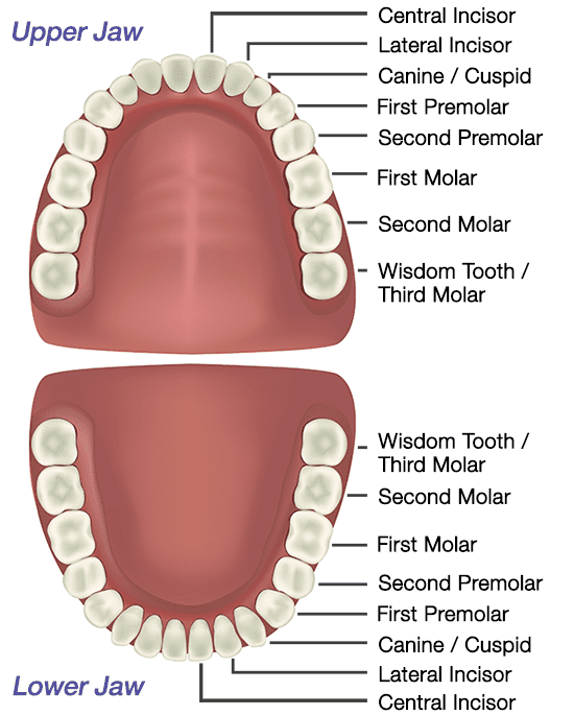
When you smile, the visible part of the tooth is called the crown. Most people refer to the crown as the “tooth” however, the crown is only one half of the tooth’s anatomy.
In this issue, we will discuss the anatomy of adult permanent teeth and demonstrate how the shape, size and appearance of a tooth determine their intended function.
The crown is only the part of the tooth that stands above the gumline. The incisors, molars, and canines all have different functions based upon their unique shape and size.
Within each crown of the tooth there are enamel, dentin, cementum and pulp. The enamel, makes up the tough outer coating responsible for protecting the internal structures. Tooth enamel is comprised of calcium phosphate minerals and is the hardest tissue in the human body. Despite being the hardest tissue, it can still be damaged. Enamel does not contain living cells and cannot repair itself. When the enamel is damaged, a dentist is needed correct the damage.
Below the enamel, you have dentin which is composed of several microscopic tiny tunnels (tubes or canals) and minerals. Dentin is nine times softer than enamel. When your tooth’s enamel gets severely damaged, your dentin becomes exposed; causing sensitivity to hot, cold, acidic or sweets. This sensitivity is a result of stimulants traveling through the tiny tunnels directly to the center of your tooth.
The cementum is the hard-connective tissue covering the tooth root that gives attachment to the periodontal ligament (PDL).
The periodontal ligament (PDL) is a system of collagen fibers that forms the joint that connects the root of the tooth to its bony socket. During periodontal disease this ligament is damaged and destroyed to some degree. With gum treatment the PDL can be restored if the damage is not too excessive.
The pulp is considered soft tissue and is located at the center of the tooth. It contains nerves, blood vessels and connective tissue. It starts from the center of the crown and extends through the roots then finally connecting to the bone for nourishment.
When Form Follows Function-
Teeth are an important part of your body. They aid in the digestive process and make clear speech possible. Each tooth has a unique anatomical form. The form of your teeth is made in such a way that it establishes its function.
Adults have 32 teeth. These include 4 wisdom teeth if they are present. Of the 32 teeth, from front to back; 8 are incisors, 4 are canines, 8 are premolars, and 12 are molars.
Incisors: Incisors are the teeth that are in the front of your mouth. The tips of these teeth are flat. They help you cut up food. They are critical to your natural smile and personality.
Canines: These are the pointy and large teeth that serve as the corner-stone of the mouth. They are good for tearing and holding food. Because of their demands their roots are the longest of all teeth.
Premolars: Premolars have 2 bumps called cusps. They are great for tearing and crushing food.
Molars: Molars are what persons refer to as the back teeth. These have multiple bumps (cusps) used for grinding and chewing food. Because of demands they are multi-rooted which add to their strong foundation. In fact, your molars can have a total bite pressure of about 5600 pounds per square inch. That’s a lot of force.
Your teeth help you bite, chew and digest food. Also, they assist with word pronunciation and speech. Each tooth consists of various anatomical parts with unique properties and functions.
Learning the basics of tooth anatomy will help you understand how their form follows function. Take care of this complex system to help you enjoy a healthy smile.
Dr. Kendal V. O. Major is Founder and CEO of Center for Specialized Dentistry which is a comprehensive family dental practice operating in Nassau and Freeport. He is the first Bahamian Specialist in gum diseases and dental implants since 1989. He also is a certified Fast braces provider. His practice is located at 89 Collins Avenue, Nassau at (242)325-5165 or [email protected]


